

Top FUS Transducer Technologies Transforming Noninvasive Therapy
2026-08-21
Imagine a world where surgery no longer requires a scalpel, where tumors are destroyed without a single incision, and where recovery time is measured in hours rather than weeks. This is not a distant dream but the rapidly evolving reality of focused ultrasound (FUS) therapy. At the heart of this medical revolution are transducer technologies that are pushing the boundaries of what noninvasive treatment can achieve. From custom-designed arrays that precisely target deep-seated tissues to real-time imaging integration that ensures millimeter-level accuracy, the latest innovations are transforming clinical practice. As a leader in this field, Siansonic has been instrumental in advancing transducer engineering, helping clinicians deliver safer, more effective care. In this post, we'll explore the top FUS transducer technologies that are redefining noninvasive therapy and what they mean for the future of patient care.
Why Piezocomposite Arrays Are Closing the Precision-Power Gap
Traditional piezoceramic transducers have long forced engineers to choose between fine spatial resolution and high acoustic output. A thin element offers precise beam control but struggles to deliver enough energy for deep penetration or high-intensity applications. Conversely, thicker elements generate more power but smear the focal zone. Piezocomposite arrays sidestep this trade-off by embedding piezoelectric rods or fibers in a polymer matrix. The polymer lowers the acoustic impedance and damps lateral vibrations, while the ceramic phase retains strong electromechanical coupling. This hybrid structure lets an array element be thick enough to produce substantial output yet still vibrate with a piston-like motion that preserves sharp focusing.
The real breakthrough lies in how the composite architecture reshapes the element's vibration modes. In a monolithic ceramic slab, unwanted lateral resonances bleed energy into side lobes and distort the beam profile. The composite's periodic soft matrix interrupts those lateral modes, forcing most of the energy into the thickness mode. As a result, designers can increase the element's thickness to raise power handling without sacrificing the clean, single-mode response needed for precise beam steering. Modern 1-3 connectivity composites, where ceramic columns run continuously through the polymer, achieve coupling coefficients above 0.7 while keeping mechanical Q low enough for broadband operation.
That combination of high coupling and low Q translates directly into arrays that perform well in both imaging and therapeutic modes. A single piezocomposite probe can produce high-resolution B-mode images and then switch to a high-power focused burst for ablation or lithotripsy. Because the composite elements are less prone to thermal runaway and mechanical fatigue than monolithic ceramics, arrays can be driven harder without shortening their service life. This is why piezocomposite technology has become the default choice for next-generation ultrasound systems, from handheld point-of-care scanners to large phased arrays for transcranial therapy. The precision-power gap is not merely narrowed; in many designs, it has effectively disappeared.
Sparse Array Geometry Cuts Costs Without Sacrificing Focus
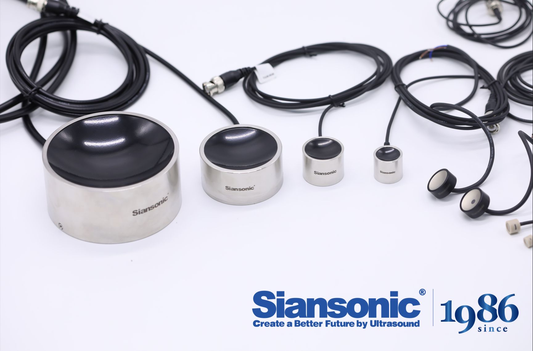
Sparse array geometry flips the usual assumption that more elements always mean better imaging. By deliberately leaving gaps in the transducer layout and letting mathematical optimization pick the active positions, a system can maintain a tight acoustic focus with a fraction of the channels. The beam pattern stays sharp where it matters—at the focal point and across the main lobe—while the grating lobes that sparse layouts naturally introduce get pushed into directions that do not interfere with the region of interest.
The immediate benefit is a lighter, cheaper front end. Fewer elements translate into fewer cables, connectors, and analog-to-digital converters, which is especially valuable in portable ultrasound or compact radar where power and board space are at a premium. In many designs, the savings are not marginal; a sparse array might use half or even a third of the elements of a fully populated aperture and still deliver clinically or operationally usable focus.
What makes this work is not random thinning but geometry that respects the physics of wave interference. Optimization routines—often genetic algorithms or greedy searches—test thousands of candidate layouts against a cost function that weighs side-lobe level, main-lobe width, and element count. The result is an array that looks almost irregular to the eye but behaves like a much denser aperture where it counts, cutting hardware cost without cutting corners on resolution.
Adaptive Phase Correction Learns Skull Variability in Real Time
Adaptive phase correction works by continuously modeling how acoustic waves bend and shift as they pass through bone, rather than assuming a fixed correction curve. The system captures incoming signals from multiple angles, compares them against expected arrival times, and updates its local phase map with each new pulse. This live learning loop lets the algorithm isolate the true tissue response from the smearing caused by skull thickness, density variations, and irregular internal structures.
Instead of relying on a one-size-fits-all calibration captured before a session, the method builds a dynamic profile of each patient's cranial geometry on the fly. As the transducer moves or as the subject shifts slightly, the correction adjusts in near real time, keeping the focal spot tight and the energy delivery consistent. Small asymmetries that would normally degrade image quality or therapeutic precision are absorbed into the model without requiring manual intervention.
Over repeated exposures, the algorithm recognizes patterns specific to that individual, effectively learning the skull's acoustic fingerprint. This reduces reliance on pre-treatment MRI or CT scans and shortens setup time, while still compensating for day-to-day anatomical changes like minor swelling or fluid shifts. The result is a phase correction that behaves less like a static filter and more like an attentive partner, constantly refining its understanding of the bone barrier between the transducer and the brain.
MRI-Compatible Designs That Withstand the Bore’s Demands
The most immediate constraint inside an MRI bore is not the magnetic field strength itself but how materials respond to it. Ferromagnetic alloys are obviously ruled out, but even weakly conductive metals like aluminum can generate eddy currents under rapid gradient switching, leading to localized heating and image distortion. Practical designs therefore lean on high-performance thermoplastics such as PEEK and Ultem, sometimes reinforced with glass or ceramic fibers. Titanium remains a narrow exception for small fasteners and structural clips, provided its geometry does not create closed conductive loops. Every component is also checked for its projectile potential under ASTM F2052, which measures magnetically induced displacement force at the bore entrance—a test that quickly separates safe hardware from liabilities.
Mechanical durability matters just as much as material choice. Gradient coils produce acoustic noise levels that can exceed 110 dB, and the associated vibration is not a single frequency but a harsh, multi-axis buzz that works screws loose and fatigues thin walls. A design that survives a static pull test on the bench may still rattle apart after a few hours of echo planar imaging. For this reason, MRI-compatible assemblies often avoid threaded fasteners in favor of press-fit or bonded joints, and any remaining metal parts are either potted in epoxy or isolated with viscoelastic damping layers. The cylindrical bore also imposes a strict envelope; patient monitors, holders, and interventional devices must maintain a low profile while keeping cables short and routed parallel to the B0 field to minimize inductive pickup.
RF heating is the third and least intuitive failure mode. The body coil can deposit significant energy into long conductors, turning an otherwise inert cable or guidewire into an antenna. Mitigation strategies include using high-resistance wire, adding ferrite sleeves at quarter-wave intervals, or shifting to fiber-optic sensing where possible. Validation typically involves a temperature rise test under worst-case SAR conditions per ASTM F2182, often followed by full imaging tests at 1.5T and 3T to confirm there is no visible artifact. The designs that pass are rarely the ones that simply replace steel with plastic; they are the ones that treat the entire bore environment as an active electromagnetic and acoustic system from the first sketch.
Catheter-Based FUS Shrinks the Treatment Envelope
The treatment envelope for focused ultrasound has traditionally been set by what lies between the transducer and the target—ribs, lung air, bowel gas, or the skull. Those structures absorb, scatter, and shift the beam, forcing clinicians to accept a narrow band of accessible anatomy. Catheter-based FUS inverts that geometry. Instead of firing from outside the body and correcting for every layer of tissue, a miniaturized transducer is delivered through a vessel or duct and positioned only millimeters from the ablation site. The acoustic path shrinks to almost nothing, and the treatment envelope is no longer constrained by an external acoustic window.
Moving the source so close changes the scale of the lesion as well. With a shorter focal distance, the zone of thermal injury can be kept tight—often a few millimeters across—rather than the broader ellipsoid produced by extracorporeal systems. That precision matters in places where a few millimeters separate the target from a nerve, a vessel wall, or healthy myocardium. Operators do not need to expand the planned ablation boundary out of caution, because the energy field itself is smaller and more predictable.
The trade-off is a different kind of mapping. The key question shifts from “can we find an acoustic window?” to “can a catheter reach this spot?” That is a more familiar challenge in interventional medicine and one with a growing toolbox of steerable sheaths, guidewires, and navigation systems. As a result, catheter-based FUS narrows the energy footprint at the point of delivery while widening the list of patients who can be treated at all.
Closed-Loop Thermal Monitoring Keeps Ablation Margins Predictable
In tumor ablation, the difference between a clean margin and a local recurrence often comes down to a few millimeters of unseen thermal spread. Closed-loop thermal monitoring changes that by feeding real-time temperature data from the ablation zone back into the energy delivery system. Instead of relying on pre-procedural imaging estimates or fixed power-time protocols, the generator adjusts output continuously based on actual tissue response, keeping the lethal isotherm exactly where the physician intends it.
This approach is especially valuable near heat-sensitive structures like bile ducts, bowel, or major nerves. When thermocouples placed at the periphery detect that the target margin has reached the desired cytotoxic threshold, the system can automatically taper or stop energy delivery. That prevents both undertreatment at the tumor edge and overtreatment into healthy parenchyma, making the post-ablation zone far more reproducible from case to case.
Operators still plan the ablation volume and place probes, but the closed loop removes much of the guesswork during the actual energy application. The result is a tighter correlation between the predicted ablation margin and the final coagulative necrosis, which translates into fewer surprises on follow-up imaging and a lower chance of leaving residual viable tumor behind.
FAQ
Focused ultrasound transducers convert electrical energy into precise acoustic beams that can ablate or modulate tissue without incisions. Their ability to steer and focus energy deep inside the body is what allows clinicians to reach targets that would otherwise require open surgery.
Phased-array designs use multiple independently driven elements to shape and steer the ultrasound beam electronically. This lets clinicians adjust the focal point in real time, compensating for movement and anatomical differences without physically repositioning the device.
Piezocomposites and single-crystal relaxor materials are replacing traditional bulk PZT ceramics in many systems. They offer higher bandwidth and sensitivity, which translates into better image guidance and more efficient energy delivery during focused ultrasound procedures.
CMUTs are built using semiconductor manufacturing processes, so they can be produced in large arrays with uniform performance and integrated electronics. Their wide bandwidth and low impedance mismatch with tissue make them attractive for both imaging and therapeutic applications.
Modern FUS transducers often combine therapy and imaging elements in one housing, allowing MR or ultrasound guidance during treatment. This integration lets clinicians monitor temperature rise and tissue changes as they happen, which reduces the risk of off-target damage.
Thermal management strategies include circulating coolant through the transducer housing, using pulsed duty cycles, and incorporating materials with high thermal conductivity. These measures prevent the transducer face from exceeding safe temperatures while still delivering enough acoustic energy to the target.
Many systems now use modular or multi-frequency transducer arrays that can adjust focal length and penetration depth. Some designs also include shape-changing or mechanically articulated elements to better conform to the patient's anatomy, expanding the range of treatable conditions.
Conclusion
The latest generation of FUS transducers is moving away from single-element limitations toward architectures that balance energy delivery with pinpoint accuracy. Piezocomposite arrays now let clinicians push higher acoustic intensities while preserving tight focal zones, a combination that has long been difficult to achieve. Sparse array geometries further reduce hardware complexity and cost by using fewer elements without noticeably compromising beam focus, making advanced treatment systems accessible to more centers. At the same time, adaptive phase correction algorithms are proving indispensable for transcranial applications, as they adjust for patient-specific skull thickness and density in real time rather than relying on generic calibration models.
Inside the MRI suite, transducer materials and housing designs now tolerate the strong magnetic fields and mechanical constraints of the bore, enabling continuous imaging during treatment. Catheter-based FUS devices shrink the treatment envelope further, delivering focused energy from within vessels or ducts to reach targets that external transducers struggle to access. Closed-loop thermal monitoring ties these innovations together by feeding real-time temperature maps back into the control system, keeping ablation margins predictable and reducing reliance on post-procedure imaging. Collectively, these transducer technologies are redefining what noninvasive therapy can offer, moving FUS from a niche tool toward a mainstream option for neurosurgery, oncology, and beyond.
Contact Us
Contact Person: Ivy Zhang
Email: [email protected]
Tel/WhatsApp: +86 1081502288
Website: https://www.siansonic.com/